11.3 The Kidneys & Osmoregulation
Essential idea:
Understandings
11.3.U1 Animals are either osmoregulators or osmoconformers.
11.3.U2 The Malpighian tubule system in insects and the kidney carry out osmoregulation and removal of nitrogenous wastes.
11.3.U3 The composition of blood in the renal artery is different from that in the renal vein.
11.3.U4 The ultrastructure of the glomerulus and Bowman’s capsule facilitate ultrafiltration.
11.3.U5 The proximal convoluted tubule selectively reabsorbs useful substances by active transport.
11.3.U6 The loop of Henle maintains hypertonic conditions in the medulla.
11.3.U7 ADH controls reabsorption of water in the collecting duct. [ADH will be used in preference to vasopressin. ]
11.3.U8 The length of the loop of Henle is positively correlated with the need for water conservation in animals.
11.3.U9 The type of nitrogenous waste in animals is correlated with evolutionary history and habitat.
Application
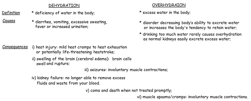
11.3 A 1 Consequences of dehydration and overhydration.
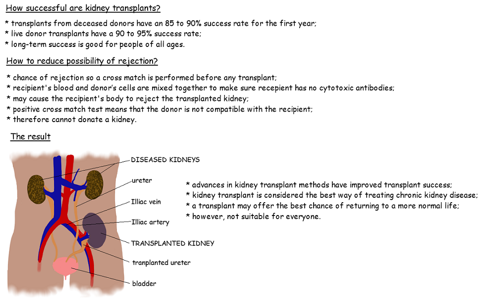
11.3 A 2 Treatment of kidney failure by hemodialysis or kidney transplant.
11.3 A 3 Blood cells, glucose, proteins and drugs are detected in urinary tests.
Skills
11.3.S1 Drawing and labelling a diagram of the human kidney.
11.3.S2 Annotation of diagrams of the nephron. [The diagram of the nephron should include glomerulus, Bowman’s capsule, proximal convoluted tubule, loop of Henle, distal convoluted tubule; the relationship between the nephron and the collecting duct should be included. ]
Essential idea:
- All animals excrete nitrogenous waste products and some animals also balance water and solute concentrations.
Understandings
11.3.U1 Animals are either osmoregulators or osmoconformers.
11.3.U2 The Malpighian tubule system in insects and the kidney carry out osmoregulation and removal of nitrogenous wastes.
11.3.U3 The composition of blood in the renal artery is different from that in the renal vein.
11.3.U4 The ultrastructure of the glomerulus and Bowman’s capsule facilitate ultrafiltration.
11.3.U5 The proximal convoluted tubule selectively reabsorbs useful substances by active transport.
11.3.U6 The loop of Henle maintains hypertonic conditions in the medulla.
11.3.U7 ADH controls reabsorption of water in the collecting duct. [ADH will be used in preference to vasopressin. ]
11.3.U8 The length of the loop of Henle is positively correlated with the need for water conservation in animals.
11.3.U9 The type of nitrogenous waste in animals is correlated with evolutionary history and habitat.
Application
11.3 A 1 Consequences of dehydration and overhydration.
11.3 A 2 Treatment of kidney failure by hemodialysis or kidney transplant.
11.3 A 3 Blood cells, glucose, proteins and drugs are detected in urinary tests.
Skills
11.3.S1 Drawing and labelling a diagram of the human kidney.
11.3.S2 Annotation of diagrams of the nephron. [The diagram of the nephron should include glomerulus, Bowman’s capsule, proximal convoluted tubule, loop of Henle, distal convoluted tubule; the relationship between the nephron and the collecting duct should be included. ]
|
|
|
|
|
|
11.3.U9 The type of nitrogenous waste in animals is correlated with evolutionary history and habitat.
Excretion is the removal from the body of the waste products of metabolic activity
There are two key functions that an excretory system performs:
Removing Nitrogenous Waste
Nitrogenous wastes are produced from the breakdown of nitrogen-containing compounds like amino acids and nucleotides
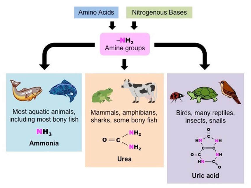
Most aquatic animals eliminate their nitrogenous wastes as ammonia (NH3)
Terrestrial animals have less access to water and hence must package nitrogenous waste in less toxic forms
Nitrogenous Waste Products
Excretion is the removal from the body of the waste products of metabolic activity
- Defecation is not considered part of excretion as feces are undigested food remnants and not metabolic waste products
There are two key functions that an excretory system performs:
- Removes nitrogenous wastes that may be toxic to the body in large concentrations
- Removes excess water to maintain a suitable osmolarity within the tissues and cells
Removing Nitrogenous Waste
Nitrogenous wastes are produced from the breakdown of nitrogen-containing compounds like amino acids and nucleotides
- Nitrogenous wastes are toxic to the organism and hence excess levels must be eliminated from the body
- The type of nitrogenous waste in animals is correlated with the evolutionary history of the animal and the habitat
Most aquatic animals eliminate their nitrogenous wastes as ammonia (NH3)
- Ammonia is highly toxic but also very water soluble and hence can be effectively flushed by animals in aquatic habitats
Terrestrial animals have less access to water and hence must package nitrogenous waste in less toxic forms
- Mammals eliminate their nitrogenous wastes as urea, which is less toxic and hence can be stored at higher concentrations
- Reptiles and birds eliminate wastes as uric acid, which requires more energy to make but is relatively non-toxic and requires even less water to flush (it is eliminated as a semi-solid paste)
Nitrogenous Waste Products
11.3.U1 Animals are either osmoregulators or osmoconformers.
Removing Excess Water
Water levels within an organism are constantly changing as a result of metabolic activity
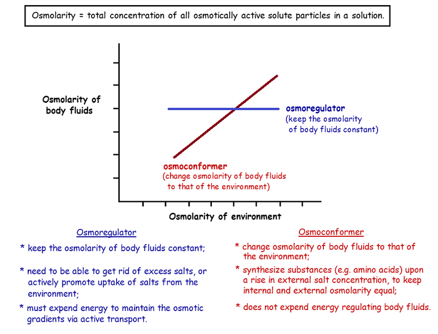
Animals may be either osmoconformers or osmoregulators according to how they manage their internal osmotic conditions:
Removing Excess Water
Water levels within an organism are constantly changing as a result of metabolic activity
- Water is produced via condensation reactions (anabolism) and is consumed during hydrolysis reactions (catabolism)
- The concentration of water within cells (osmolarity) will impact tissue viability (i.e. governs osmotic pressure within cells)
Animals may be either osmoconformers or osmoregulators according to how they manage their internal osmotic conditions:
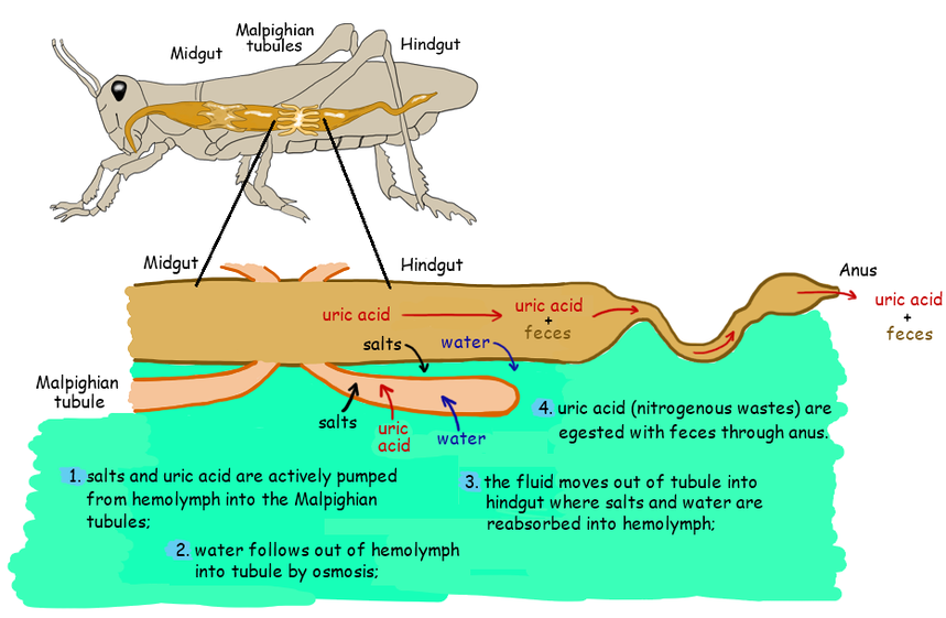
11.3.U2 The Malpighian tubule system in insects and the kidney carry out osmoregulation and removal of nitrogenous wastes.
All animals possess a specialised excretory system for osmoregulation and the removal of nitrogenous wastes
All animals possess a specialised excretory system for osmoregulation and the removal of nitrogenous wastes
- In mammals, the excretory system (kidneys) is separate from the digestive system of the animal
- In insects, the excretory system (Malpighian tubules) connects to the digestive system of the animal
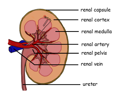
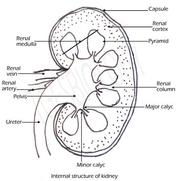
11.3.S1 Drawing and labeling a diagram of the human kidney.
The kidney functions as the blood’s filtration and water balancing system – it removes metabolic wastes for excretion
The kidney functions as the blood’s filtration and water balancing system – it removes metabolic wastes for excretion
- Blood enters the kidneys via the renal artery and exits the kidneys via the renal vein
- Blood is filtered by specialised structures called nephrons which produce urine
- The urine is transported from the kidneys via the ureter, where it is stored by the bladder prior to excretion
|
|
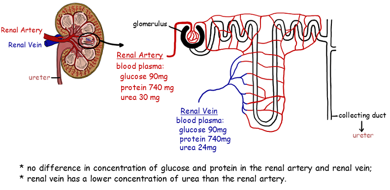
11.3.U3 The composition of blood in the renal artery is different from that in the renal vein.
The kidney contains specialised structures called nephrons which function to filter the blood and eliminate wastes
Blood in the renal vein (i.e. after the kidney) will have:
The kidney contains specialised structures called nephrons which function to filter the blood and eliminate wastes
- Consequently, the composition of blood entering the kidney (via renal artery) differs to that exiting the kidney (via renal vein)
Blood in the renal vein (i.e. after the kidney) will have:
- Less urea (large amounts of urea is removed via the nephrons to form urine)
- Less water and solutes / ions (amount removed will depend on the hydration status of the individual)
- Less glucose and oxygen (not eliminated, but used by the kidney to generate energy and fuel metabolic reactions)
- More carbon dioxide (produced by the kidneys as a by-product of metabolic reactions)
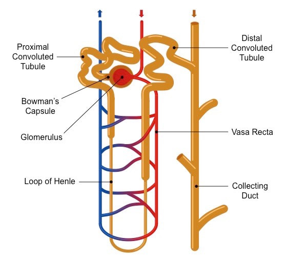
11.3.S2 Annotation of diagrams of the nephron. [The diagram of the nephron should include glomerulus, Bowman’s capsule, proximal convoluted tubule, loop of Henle, distal convoluted tubule; the relationship between the nephron and the collecting duct should be included. ]
The nephron is the functional unit of the kidney, with each nephron being comprised of the following components:
The blood to be filtered enters the Bowman’s capsule via an afferent arteriole and leaves the capsule via an efferent arteriole
Each nephron connects to a collecting duct (via the distal convoluted tubule), which feed into the renal pelvis
Structure of a Nephron
The nephron is the functional unit of the kidney, with each nephron being comprised of the following components:
- Bowman’s capsule – first part of the nephron where blood is initially filtered (to form filtrate)
- Proximal convoluted tubule – folded structure connected to the Bowman’s capsule where selective reabsorption occurs
- Loop of Henle – a selectively permeable loop that descends into the medulla and establishes a salt gradient
- Distal convoluted tubule – a folded structure connected to the loop of Henle where further selective reabsorption occurs
The blood to be filtered enters the Bowman’s capsule via an afferent arteriole and leaves the capsule via an efferent arteriole
- Within the Bowman’s capsule, the blood is filtered at a capillary tuft called the glomerulus
- The efferent arteriole forms a blood network called the vasa recta that reabsorbs components of the filtrate from the nephron
Each nephron connects to a collecting duct (via the distal convoluted tubule), which feed into the renal pelvis
- The collecting ducts are shared by nephrons and hence are not technically considered to be part of a single nephron
Structure of a Nephron
Function of the Nephron
|
|
|
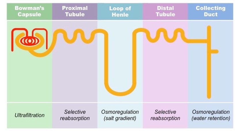
Nephron Function
Nephrons filter blood and then reabsorb useful materials from the filtrate before eliminating the remainder as urine
This process occurs over three key stages:
Relationship between Nephron Structure and Function
Nephrons filter blood and then reabsorb useful materials from the filtrate before eliminating the remainder as urine
This process occurs over three key stages:
- Ultrafiltration – Blood is filtered out of the glomerulus at the Bowman’s capsule to form filtrate
- Selective reabsorption – Usable materials are reabsorbed in convoluted tubules (both proximal and distal)
- Osmoregulation – The loop of Henle establishes a salt gradient, which draws water out of the collecting duct
Relationship between Nephron Structure and Function
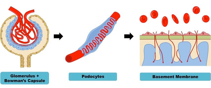
11.3.U4 The ultrastructure of the glomerulus and Bowman’s capsule facilitate ultrafiltration.
Ultrafiltration is the first of three processes by which metabolic wastes are separated from the blood and urine is formed
Ultrafiltration is the first of three processes by which metabolic wastes are separated from the blood and urine is formed
- It is the non-specific filtration of the blood under high pressure and occurs in the Bowman’s capsule of the nephron
- As the blood moves into the kidney via afferent arterioles it enters a knot-like capillary tuft called a glomerulus
- This glomerulus is encapsulated by the Bowman’s capsule, which is comprised of an inner surface of cells called podocytes
- Podocytes have cellular extensions called pedicels that wrap around the blood vessels of the glomerulus
- Between the podocytes and the glomerulus is a glycoprotein matrix called the basement membrane that filters the blood
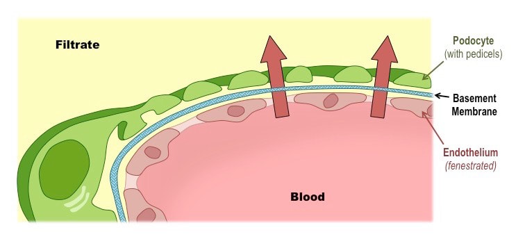
Basement Membrane
Blood is filtered by a mesh called the basement membrane, which lies between the glomerulus and Bowman’s capsule
The basement membrane is size-selective and restricts the passage of blood cells and large proteins
Blood is filtered by a mesh called the basement membrane, which lies between the glomerulus and Bowman’s capsule
- Glomerular blood vessels are fenestrated (have pores) which means blood can freely exit the glomerulus
- The podocytes of the Bowman’s capsule have gaps between their pedicels, allowing for fluid to move freely into the nephron
- Consequently, the basement membrane functions as the sole filtration barrier within the nephron
The basement membrane is size-selective and restricts the passage of blood cells and large proteins
- Hence when the blood is filtered, the filtrate formed does not contain any blood cells, platelets or plasma proteins
Hydrostatic Pressure
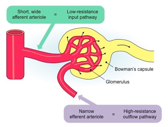
Ultrafiltration involves blood being forced at high pressure against the basement membrane, optimising filtration
Ultrafiltration involves blood being forced at high pressure against the basement membrane, optimising filtration
- This high hydrostatic pressure is created in the glomerulus by having a wide afferent arteriole and a narrow efferent arteriole
- This means it is easy for blood to enter the glomerulus, but difficult for it to exit – increasing pressure within the glomerulus
- Additionally, the glomerulus forms extensive narrow branches, which increases the surface area available for filtration
- The net pressure gradient within the glomerulus forces blood to move into the capsule space (forming filtrate)
11.3.U5 The proximal convoluted tubule selectively reabsorbs useful substances by active transport.

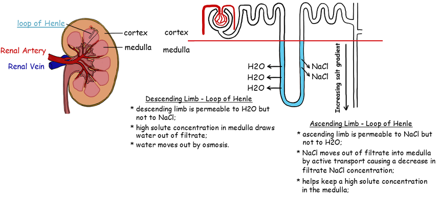
11.3.U6 The loop of Henle maintains hypertonic conditions in the medulla.
Osmoregulation is the third of three processes by which blood is filtered and urine is formed
Osmoregulation occurs in the medulla of the kidney and involves two key events:
Osmoregulation is the third of three processes by which blood is filtered and urine is formed
- Osmoregulation is the control of the water balance of the blood, tissue or cytoplasm of a living organism
Osmoregulation occurs in the medulla of the kidney and involves two key events:
- The loop of Henle establishes a salt gradient (hypertonicity) in the medulla
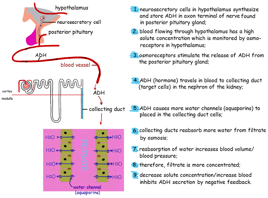
- Anti-diuretic hormone (ADH) regulates the level of water reabsorption in the collecting duct
11.3.U7 ADH controls reabsorption of water in the collecting duct. [ADH will be used in preference to vasopressin. ]
Collecting Duct
Collecting Duct
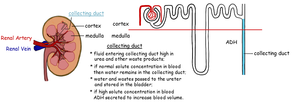
Water Reabsorption
Remember: ADH is produced when you Are DeHydrated
Remember: ADH is produced when you Are DeHydrated
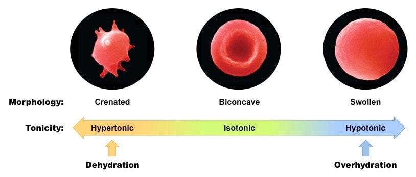
11.3 A 1 Consequences of dehydration and overhydration.
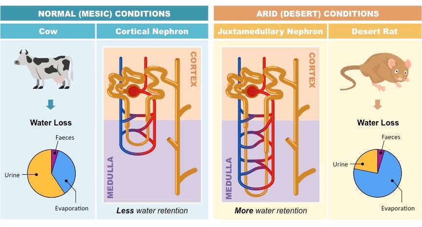
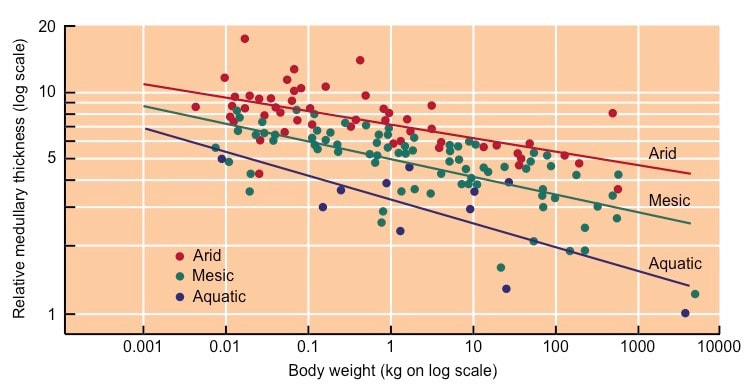
11.3.U8 The length of the loop of Henle is positively correlated with the need for water conservation in animal
The length of the loop of Henle is positively correlated with the degree of water conservation in animals
Comparison of Nephrons in Mesic and Desert Animals
The length of the loop of Henle is positively correlated with the degree of water conservation in animals
- Animals living in moist environments have short loops of Henle that don’t descend deeply into the medulla (cortical nephrons)
- Animals living in arid environments have long loops of Henle that descend deeply into the medulla (juxtamedullary nephrons)
Comparison of Nephrons in Mesic and Desert Animals
11.3 A 3 Blood cells, glucose, proteins and drugs are detected in urinary tests.
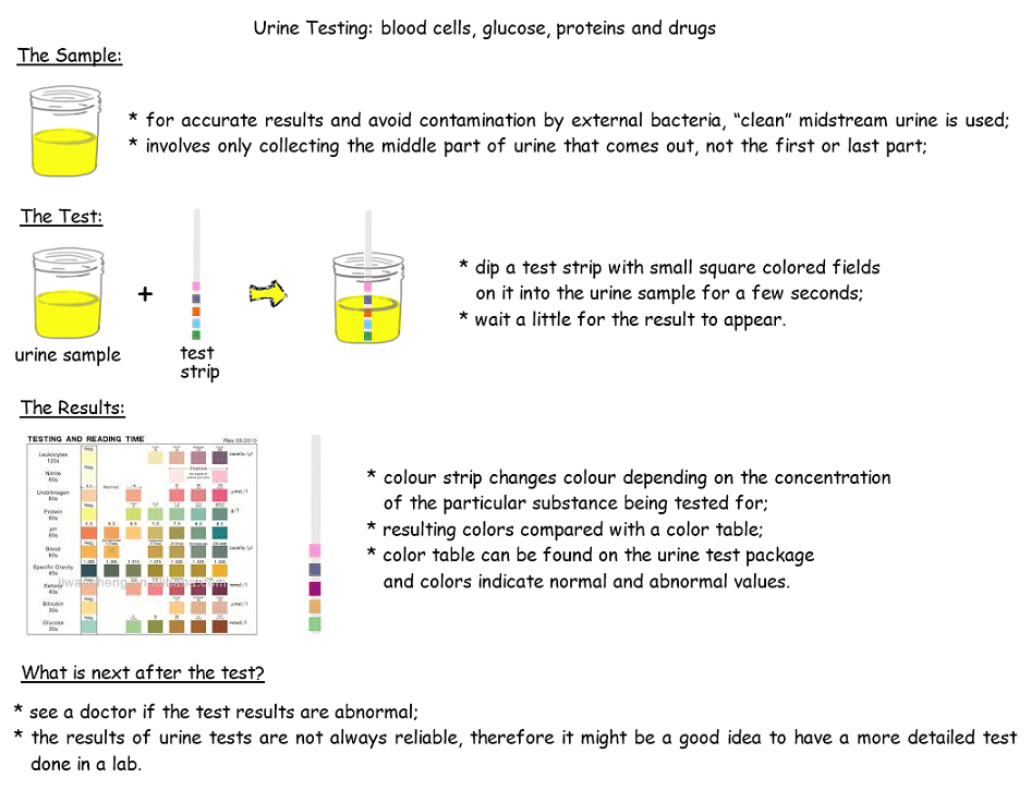
Kidneys prevent the excretion of blood cells and proteins (during ultrafiltration), as well as glucose (selective reabsorption)
Glucose: The presence of glucose in urine is a common indicator of diabetes (high blood glucose = incomplete reabsorption)
Proteins: High quantities of protein in urine may indicate disease (e.g. PKU) or hormonal conditions (e.g. hCG = pregnancy)
Blood cells: The presence of blood in urine can indicate a variety of diseases, including certain infections and cancer
Drugs / toxins: Many drugs pass through the body into urine and can be detected (e.g. performance enhancing drugs)
- Hence the presence of these materials in urine can be used as an indicator of disease
Glucose: The presence of glucose in urine is a common indicator of diabetes (high blood glucose = incomplete reabsorption)
Proteins: High quantities of protein in urine may indicate disease (e.g. PKU) or hormonal conditions (e.g. hCG = pregnancy)
Blood cells: The presence of blood in urine can indicate a variety of diseases, including certain infections and cancer
Drugs / toxins: Many drugs pass through the body into urine and can be detected (e.g. performance enhancing drugs)
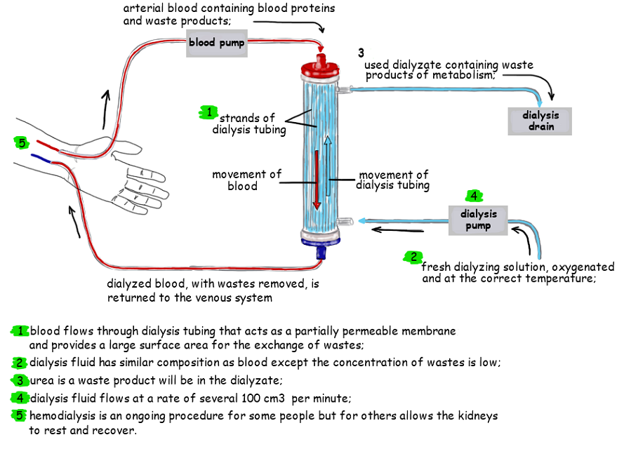
11.3 A 2 Treatment of kidney failure by hemodialysis or kidney transplant.
Hemodialysis
Kidney dialysis involves the external filtering of blood in order to remove metabolic wastes in patients with kidney failure
Blood is removed and pumped through a dialyzer, which has two key functions that are common to biological membranes:
Kidney dialysis treatments typically last about 4 hours and occur 3 times a week – these treatments can be effective for years
Hemodialysis
Kidney dialysis involves the external filtering of blood in order to remove metabolic wastes in patients with kidney failure
Blood is removed and pumped through a dialyzer, which has two key functions that are common to biological membranes:
- It contains a porous membrane that is semi-permeable (restricts passage of certain materials)
- It introduces fresh dialysis fluid and removes wastes to maintain an appropriate concentration gradient
Kidney dialysis treatments typically last about 4 hours and occur 3 times a week – these treatments can be effective for years