2.6 Structure of DNA and RNA
Essential Idea:
The structure of DNA allows efficient storage of genetic information
Understandings:
2.6.U1: The nucleic acids DNA and RNA are polymers of nucleotides.
2.6.U2: DNA differs from RNA in the number of strands present, the base composition and the type of pentose.
2.6.U3: DNA is double helix made of two antiparallel strands of nucleotides linked by hydrogen bonding between complementary base pairs.
Applications:
2.6.A1: Crick and Watson’s elucidation of the structure of DNA using model making.
Skills:
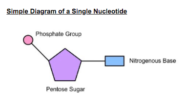
2.6.S1: Drawing simple diagrams of the structure of single nucleotides of DNA and RNA, using circles, pentagons, and rectangles to represent phosphates, pentoses and bases.
The structure of DNA allows efficient storage of genetic information
Understandings:
2.6.U1: The nucleic acids DNA and RNA are polymers of nucleotides.
2.6.U2: DNA differs from RNA in the number of strands present, the base composition and the type of pentose.
2.6.U3: DNA is double helix made of two antiparallel strands of nucleotides linked by hydrogen bonding between complementary base pairs.
Applications:
2.6.A1: Crick and Watson’s elucidation of the structure of DNA using model making.
Skills:
2.6.S1: Drawing simple diagrams of the structure of single nucleotides of DNA and RNA, using circles, pentagons, and rectangles to represent phosphates, pentoses and bases.
DNA Structure
Understandings:
2.6.U1: The nucleic acids DNA and RNA are polymers of nucleotides.
Nucleic acids are the genetic material of the cell and are composed of recurring monomeric units called nucleotides
Each nucleotide is comprised of three principal components:
Both the phosphate group and nitrogenous base are attached to the central pentose sugar
2.6.U1: The nucleic acids DNA and RNA are polymers of nucleotides.
Nucleic acids are the genetic material of the cell and are composed of recurring monomeric units called nucleotides
Each nucleotide is comprised of three principal components:
- 5-carbon pentose sugar (pentagon)
- Phosphate group (circle)
- Nitrogenous base (rectangle)
Both the phosphate group and nitrogenous base are attached to the central pentose sugar
- The nitrogenous base is attached to the 1’– carbon atom (right point)
- The phosphate base is attached to the 5’– carbon atom (left point)
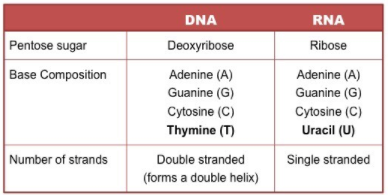
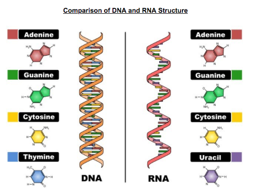
DNA versus RNA
There are two types of nucleic acids present in cells – DNA and RNA
2.6.S1: Drawing simple diagrams of the structure of single nucleotides of DNA and RNA, using circles, pentagons, and rectangles to represent phosphates, pentoses and bases.
- DNA (deoxyribonucleic acid) is a more stable double stranded form that stores the genetic blueprint for cells
- RNA (ribonucleic acid) is a more versatile single stranded form that transfers the genetic information for decoding
2.6.S1: Drawing simple diagrams of the structure of single nucleotides of DNA and RNA, using circles, pentagons, and rectangles to represent phosphates, pentoses and bases.
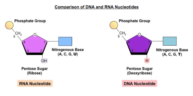
2.6.U2: DNA differs from RNA in the number of strands present, the base composition and the type of pentose.
DNA and RNA are both polymers of nucleotides, however differ in a few key structural aspects:
Differences between DNA and RNA
DNA and RNA are both polymers of nucleotides, however differ in a few key structural aspects:
- Number of strands present
- Composition of nitrogenous bases
- Type of pentose sugar
Differences between DNA and RNA
2.6/7.1 - Structure of DNA and RNA Powerpoint
From Nucleotides Into Polymers
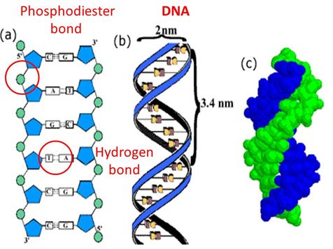
2.6.U3: DNA is double helix made of two antiparallel strands of nucleotides linked by hydrogen bonding between complementary base pairs.
Phosphate-Sugar Backbone
Nucleotides are linked together by covalent bonds between phosphate of one nucleotide and sugar of next. These monomers that are linked through phosphodiester bonds become the phosphate-sugar backbone of nucleic acids. Nitrogenous bases extending from this phosphate-sugar backbone like teeth of a comb.
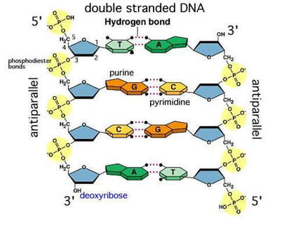
The Nucleic Acid Ladder
Hydrogen bonds form between specific bases of two nucleic acid chains, forming a stable, double-stranded DNA molecule. The structure is analogous to a ladder, with the two deoxyribose-phosphate chains as side rails and the base pairs, linked by hydrogen bonds, forming the rungs.
Phosphate-Sugar Backbone
Nucleotides are linked together by covalent bonds between phosphate of one nucleotide and sugar of next. These monomers that are linked through phosphodiester bonds become the phosphate-sugar backbone of nucleic acids. Nitrogenous bases extending from this phosphate-sugar backbone like teeth of a comb.
The Nucleic Acid Ladder
Hydrogen bonds form between specific bases of two nucleic acid chains, forming a stable, double-stranded DNA molecule. The structure is analogous to a ladder, with the two deoxyribose-phosphate chains as side rails and the base pairs, linked by hydrogen bonds, forming the rungs.
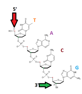
Antiparallel Strands
Notice that in the two figures above, the two strands of a DNA molecule are antiparallel, that is, they run in different directions. The side of the chain on the left begins with a free phosphate group at the top and ends with a sugar molecule at the bottom. In contrast, the complementary chain on the right begins at the top with a sugar molecule and ends at the bottom with a phosphate group.
Notice that in the two figures above, the two strands of a DNA molecule are antiparallel, that is, they run in different directions. The side of the chain on the left begins with a free phosphate group at the top and ends with a sugar molecule at the bottom. In contrast, the complementary chain on the right begins at the top with a sugar molecule and ends at the bottom with a phosphate group.
- 5 PRIME = The phosphate end is called the 5' end because the fifth carbon is closest to the phosphate group.
- 3 PRIME = The sugar end is called the 3' end because the third carbon is closest to the sugar.
|
|
Nitrogenous Bases
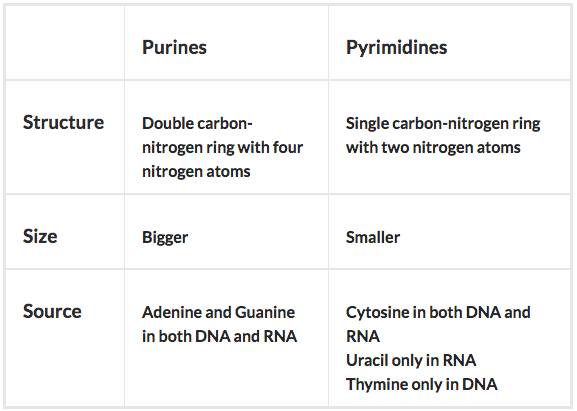
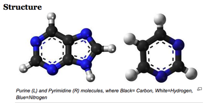
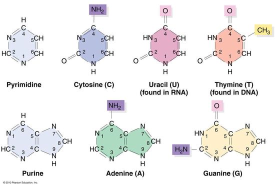
There are two types of bases: Purines and Pyrimidines. See the differences in structure and size below.
|
|
There are two main types of purines: Adenine and Guanine. Both of these occur in both DNA and RNA.
There are three main types of pyrimidines, however only one of them exists in both DNA and RNA: Cytosine. The other two are Uracil, which is RNA exclusive, and Thymine, which is DNA exclusive.
One strategy that may help you remember this is to think of pyrimidines like pyramids that have sharp and pointy tops. So sharp and pointy in fact, that they might CUT (Cytosine, Uracil, Thymine) you.
Which purines pair with which pyrimidines is always constant, as is the number of hydrogen bonds between them:
• ADENINE pairs with THYMINE (A::T) with two hydrogen bonds
• GUANINE pairs with CYTOSINE (G:::C) with three hydrogen bonds
Important Figures in the Discovery of DNA Structure
Alfred Hershey & Martha Chase
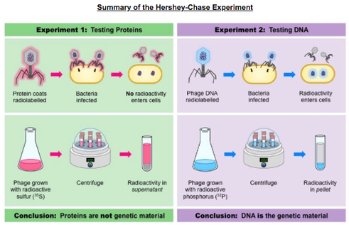
7.1.S1: Analysis of results of the Hershey and Chase experiment providing evidence that DNA is the genetic material.
In the mid-twentieth century, scientists were still unsure as to whether DNA or protein was the genetic material of the cell. It was known that some viruses consisted solely of DNA and a protein coat and could transfer their genetic material into hosts. In 1952, Alfred Hershey and Martha Chase conducted a series of experiments to prove that DNA was the genetic material:
Viruses (T2 bacteriophage) were grown in one of two isotopic mediums in order to radioactively label a specific viral component
- Viruses grown in radioactive sulfur (35S) had radiolabelled proteins (sulfur is present in proteins but not DNA)
- Viruses grown in radioactive phosphorus (32P) had radiolabeled DNA (phosphorus is present in DNA but not proteins)
The bacterial pellet was found to be radioactive when infected by the 32P–viruses (DNA) but not the 35S–viruses (protein). This demonstrated that DNA, not protein, was the genetic material because DNA was transferred to the bacteria
Rosalind Franklin & Maurice Wilkins
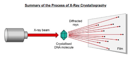
7.1.A1: Rosalind Franklin and Maurice Wilkins’ investigation of DNA structures by X-ray.
7.1 NOS Making careful observations- Rosalind Franklin’s X-ray diffraction provided crucial evidence that DNA is a double helix.
Rosalind Franklin and Maurice Wilkins used a method of X-ray diffraction to investigate the structure of DNA:
- DNA was purified and then fibres were stretched in a thin glass tube (to make most of the strands parallel)
- The DNA was targeted by a X-ray beam, which was diffracted when it contacted an atom
- The scattering pattern of the X-ray was recorded on a film and used to elucidate details of molecular structure
From the scattering pattern produced by a DNA molecule, certain inferences could be made about its structure:
Composition: DNA is a double stranded molecule
- Orientation: Nitrogenous bases are closely packed together on the inside and phosphates form an outer backbone
- Shape: The DNA molecule twists at regular intervals (every 34 Angstrom) to form a helix (two strands = double helix)
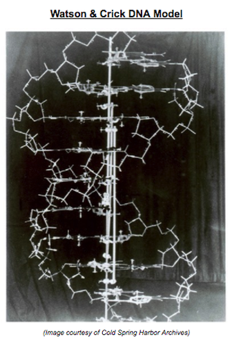
Francis Crick & James Watson
2.6.A1: Crick and Watson’s elucidation of the structure of DNA using model making.
Using trial and error, Watson and Crick were able to assemble a DNA model that demonstrated the following:
- DNA strands are antiparallel and form a double helix
As Watson and Crick’s model building was based on trial and error, a number of early models possessed faults:
2.6.A1: Crick and Watson’s elucidation of the structure of DNA using model making.
Using trial and error, Watson and Crick were able to assemble a DNA model that demonstrated the following:
- DNA strands are antiparallel and form a double helix
- - DNA strands pair via complementary base pairing (A = T ; C Ξ G)
- - Outer edges of bases remain exposed (allows access to replicative and transcriptional proteins)
As Watson and Crick’s model building was based on trial and error, a number of early models possessed faults:
- - The first model generated was a triple helix
- - Early models had bases on the outside and sugar-phosphate residues in the centre
- - Nitrogenous bases were not initially configured correctly and hence did not demonstrate complementarity