D.4: FUNCTION OF THE HEART
Essential idea:
Nature of science:
- Internal and external factors influence heart function.
Nature of science:
- Developments in scientific research followed improvements in apparatus or instrumentation—the invention of the stethoscope led to improved knowledge of the workings of the heart. (1.8)
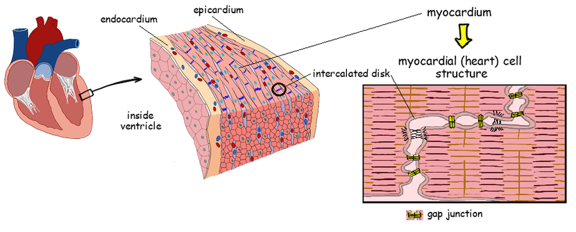 Image from Ibbiologyhelp.com
Understandings:
D.4 U 1 Structure of cardiac muscle cells allows propagation of stimuli through the heart wall.
How is cardiac muscle different from other muscle?
The heart is composed of cardiac muscle cells which have specialised features that relate to their function:
D.4 U 1 Structure of cardiac muscle cells allows propagation of stimuli through the heart wall.
How is cardiac muscle different from other muscle?
The heart is composed of cardiac muscle cells which have specialised features that relate to their function:
- Cardiac muscle cells contract without stimulation by the central nervous system (contraction is myogenic)
- Cardiac muscle cells are branched, allowing for faster signal propagation and contraction in three dimensions
- Cardiac muscles cells are not fused together, but are connected by gap junctions at intercalated discs
- Cardiac muscle cells have more mitochondria, as they are more reliant on aerobic respiration than skeletal muscle.
- Cardiac muscle has a longer period of contraction and refraction, which is needed to maintain a viable heart beat
- The heart tissue does not become fatigued (unlike skeletal muscle), allowing for continuous, life long contractions
- The interconnected network of cells is separated between atria and ventricles, allowing them to contract separately
MYOGENIC MUSCLE: muscle that has the ability to contract without receiving impulses from nerves. The contraction originates from the muscle itself.
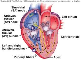
D.4 U2 Signals from the sinoatrial node that cause contraction cannot pass directly from atria to ventricles.
What stimulates the heart to change its rate of contraction?
Cardiac muscle cells are not fused together but are instead connected via gap junctions at intercalated discs
Cardiac muscle cells are not fused together but are instead connected via gap junctions at intercalated discs
- This means that while electrical signals can pass between cells, each cell is capable of independent contraction
- The coordinated contraction of cardiac muscle cells is controlled by specialised autorhythmic cells (‘pace makers’)
Atrial Contraction
Within the wall of the right atrium is a specialised cluster of cardiomyocytes which directs the contraction of heart tissue
The atria and ventricles of the heart are separated by a fibrous cardiac skeleton composed of connective tissue
Within the wall of the right atrium is a specialised cluster of cardiomyocytes which directs the contraction of heart tissue
- This cluster of cells is collectively called the sinoatrial node (SA node or SAN)
- The sinoatrial node acts as a primary pacemaker, controlling the rate at which the heart beats (i.e. pace 'making’)
- It sends out electrical signals which are propagated throughout the entire atria via gap junctions in the intercalated discs
- In response, the cardiac muscle within the atrial walls contract simultaneously (atrial systole)
The atria and ventricles of the heart are separated by a fibrous cardiac skeleton composed of connective tissue
- This connective tissue functions to anchor the heart valves in place and cannot conduct electrical signals
- The signals from the sinoatrial node must instead be relayed through a second node located within this cardiac skeleton
- This second node is called the atrioventricular node (or AV node) and separates atrial and ventricular contractions
- The AV node propagates electrical signals more slowly than the SA node, creating a delay in the passing on of the signal
Overview of Atrial Contraction/Systole
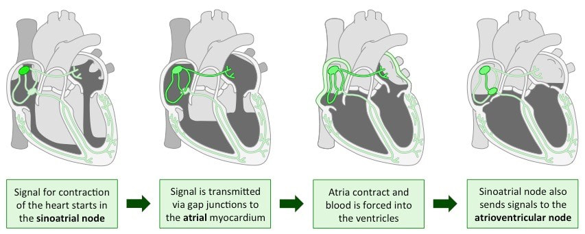
Image from Bioninja