Karyotyping is the process by which chromosomes are organised and visualised for inspection
Cells are harvested from the foetus before being chemically induced to undertake cell division (so chromosomes are visible)
Finally, chromosomes are stained and photographed, before being organised according to structure
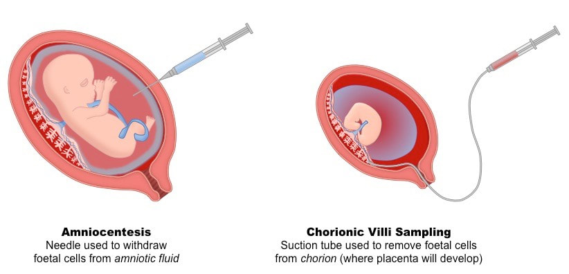
Two procedures for obtaining fetal cells for production of a karyotype:
Amniocentesis
A needle is inserted through the abdomen into the uterus. Amniotic fluid is withdrawn, which contains cells from the fetus that can be used in karyotyping. A risk is uterine contractions.
Chorionic Villus Sampling
A thin tube is inserted into the uterus via the vagina and cervix. A small sample of placenta is removed, which contains fetal cells that can be used in karyotyping. A risk is bleeding.
- Karyotyping is typically used to determine the gender of an unborn child and test for chromosomal abnormalities
Cells are harvested from the foetus before being chemically induced to undertake cell division (so chromosomes are visible)
- The stage during which mitosis is arrested will determine whether chromosomes appear with sister chromatids
Finally, chromosomes are stained and photographed, before being organised according to structure
- The visual profile generated is called a karyogram
Two procedures for obtaining fetal cells for production of a karyotype:
Amniocentesis
A needle is inserted through the abdomen into the uterus. Amniotic fluid is withdrawn, which contains cells from the fetus that can be used in karyotyping. A risk is uterine contractions.
Chorionic Villus Sampling
A thin tube is inserted into the uterus via the vagina and cervix. A small sample of placenta is removed, which contains fetal cells that can be used in karyotyping. A risk is bleeding.